What Is Interventional Radiology?
Interventional Radiology is one of the fastest-growing fields of modern medicine and has become increasingly recognized in recent years. It is a subspecialty of radiology that focuses on performing minimally invasive treatments using imaging guidance. Historically, radiology dealt mainly with diagnosis, whereas interventional radiology now provides therapeutic solutions.
How Are Interventional Radiology Procedures Performed?
Using imaging-guided, minimally invasive techniques, interventional radiology procedures are performed through the skin to effectively treat a wide range of clinical conditions. Compared to traditional surgery, these procedures are typically less invasive, cause less pain, involve fewer risks, and require shorter recovery times.
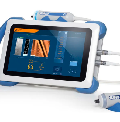
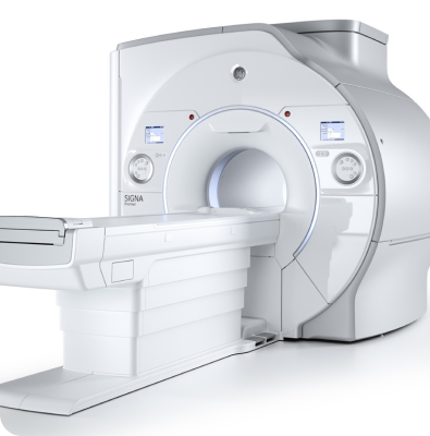
Procedures are planned in advance based on existing imaging studies. Under local or general anesthesia, treatment is performed under imaging guidance — either through the skin or via blood vessels — using catheters, guidewires, and other specialized instruments. Digital Subtraction Angiography (DSA), fluoroscopy, ultrasonography, computed tomography (CT), and magnetic resonance imaging (MRI) are the primary imaging modalities used in interventional radiology.
In Which Diseases Is Interventional Radiology Used?
Interventional radiology procedures are divided into three main categories: vascular (related to blood vessels), oncological (performed in cancer patients), and non-vascular (unrelated to blood vessels).
1) Vascular Interventional Procedures
• The most common procedure is diagnostic angiography, in which contrast material is injected into the vessel through a catheter to visualize and record images. Angiographies of the brain, leg arteries, and abdominal organs are of great importance for treatment planning.
• When vascular narrowing, blockage, clot formation, malformations, or vascular tumors are detected, interventional procedures can be used for treatment:
- Balloon angioplasty and/or stent placement for narrowing or occlusion (PTA – stenting)
- Clot removal or dissolution (thrombectomy – thrombolysis)
- Embolization to block feeding arteries of malformations or tumors
Stents are cylindrical metal mesh structures used to expand narrowed or blocked vessels. They are produced in various diameters, lengths, and designs for use in different vascular regions. Today, narrow and long balloons designed for below-the-knee arteries allow for successful treatment of lower limb occlusions.
• The main artery exiting the heart is called the aorta, and its enlargement is known as an aortic aneurysm. While these were traditionally treated through open surgery, today they can be repaired using covered stent grafts inserted endovascularly (through the vessel).
• An important sub-branch of interventional radiology is Interventional Neuroradiology, which focuses on endovascular (intravascular) treatment of brain vessel diseases:
- Narrowing or blockage in the carotid arteries (neck vessels) can be treated using stents, balloons, and specialized filters, restoring vessel patency and preventing stroke.
- In cases of acute stroke caused by blood clots blocking brain arteries, interventional neuroradiologists can remove or dissolve the clot to prevent or minimize neurological damage.
- Brain aneurysms (vascular bulges) and arteriovenous malformations (AVMs) that can cause hemorrhages can also be treated via endovascular techniques, reaching the vessels through a minimally invasive route.
- Highly vascular brain tumors may also be embolized before surgery to reduce intraoperative bleeding.
• Venous disorders:
- Diagnostic evaluation
- Deep vein thrombosis (DVT) can be treated using catheter-directed thrombolysis to dissolve clots and restore venous flow. Early intervention can result in complete resolution of the clot.
- Varicose veins:
o Treated with sclerotherapy
o Treated using laser ablation under ultrasound guidance
These procedures are performed on an outpatient basis without the need for surgery.
• Placement of hemodialysis catheters is one of the most common interventional procedures for patients requiring high-volume blood exchange. When performed under image guidance by interventional radiologists, success rates increase and complications are minimized.
• Dialysis fistulas are vital for patients undergoing hemodialysis. Over time, fistulas may develop narrowing, clots, or blockages, all of which can be effectively treated using interventional radiologic techniques, extending the lifespan of the fistula.
2) Oncological Interventional Procedures
• Placement of port catheters facilitates intravenous medication delivery, particularly in chemotherapy patients.
• Chemoembolization and Radioembolization are specialized embolization techniques primarily used in liver cancer treatment. In chemoembolization, chemotherapeutic drugs and embolic agents are delivered directly into the arteries feeding the tumor. In radioembolization, radioactive materials are used instead to deliver internal radiation therapy to the tumor.
• Radiofrequency (RFA) or Microwave Ablation techniques involve inserting a probe into the tumor under imaging guidance and applying heat to destroy the tumor tissue. These are especially effective for treating liver tumors.
3) Non-Vascular Interventional Procedures
• Biopsies: Diagnostic tissue sampling procedures performed under imaging guidance (ultrasound, CT, or MRI). Samples can be taken from organs such as the thyroid, prostate, liver, lung, kidney, and pancreas. A fine needle is inserted into the target area to obtain tissue for pathological examination, allowing definitive diagnosis in many conditions.
• Drainage procedures: Used to treat abscesses or cysts by draining fluid collections under imaging guidance.
• Nephrostomy: Placement of a catheter into the kidney’s collecting system to allow urine drainage.
• Percutaneous Transhepatic Cholangiography (PTC): A procedure in which contrast material is injected through a fine needle inserted into the liver to visualize the bile ducts.
• Percutaneous Biliary Drainage: Following visualization of the bile ducts, a drainage catheter is placed to allow bile to flow externally.
• Stent placement in bile duct obstructions to restore biliary drainage.
• Drainage of renal cysts and treatment of hydatid cysts can also be performed under imaging guidance.