PET/CT
Oncological Diagnosis and Planning
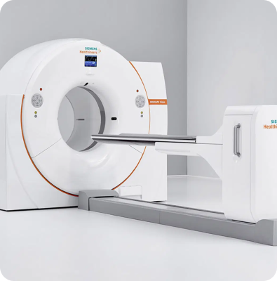
PET/CT (Positron Emission Tomography – Computed Tomography) is a hybrid imaging technology that combines nuclear medicine and radiology. By simultaneously visualizing both the structural features of organs and their metabolic activity at the cellular level, it plays a vital role in cancer diagnosis and treatment planning. PET/CT not only helps in detecting existing diseases but also allows physicians to assess treatment response and effectiveness during the therapeutic process.
Technological Infrastructure and Operation
PET/CT integrates two imaging modalities into a single system:
- PET (Positron Emission Tomography): A radioactive glucose analog, FDG (Fluorodeoxyglucose), is injected into the body. Since cancer cells consume more energy than normal cells, they absorb more FDG, making metabolically active areas visible.
- CT (Computed Tomography): Provides detailed imaging of the body’s anatomical structures.
By overlaying the PET and CT images, physicians can accurately determine both the location and extent of disease. This integrated approach aids not only in detecting cancerous tissues but also in identifying infections and inflammatory processes that exhibit high metabolic activity.
Advantages
- Enables early detection of cancer while it is still in its initial stages
- Allows accurate staging, enabling personalized treatment planning
- Helps evaluate the response to treatment, guiding adjustments in therapy
- Provides valuable insights for surgical and radiotherapy planning
- Can also be used to evaluate non-cancerous conditions such as infections and certain neurological diseases
Applications
PET/CT is widely used in several medical specialties:
- Oncology: Tumor diagnosis, staging, and monitoring treatment response
- Neurology: Identification of Alzheimer’s disease and epileptic foci
- Cardiology: Assessment of myocardial viability
- Infection and Inflammation: Detection of infection sites and inflammatory processes

Your Health
In Trusted Hands
The New Güven Health Group Mobile App Is Now Available!
Downloading our app is easy! Choose the link suitable for your operating system and start simplifying your life right away.