EBUS (Endobronşiyal Ultrasonografi)
Bronchial and Mediastinal Evaluation
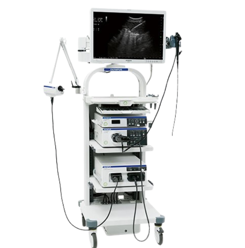
EBUS (Endobronchial Ultrasound) is an advanced imaging and diagnostic technique used for examining lymph nodes and masses located in the lungs and mediastinum (chest cavity). Combining bronchoscopy and ultrasound technologies, this method plays a vital role in the diagnosis and staging of lung cancer. Being minimally invasive, it provides highly accurate results without the need for surgical intervention.
Technological Infrastructure and Application
The EBUS device operates through a special bronchoscope equipped with an ultrasound probe at its tip. During the procedure, the bronchoscope is advanced through the mouth or nose into the trachea, allowing real-time ultrasound visualization of tissues beyond the bronchial wall.
Simultaneously, fine-needle aspiration biopsy (EBUS-TBNA) can be performed, enabling direct sampling from suspicious lymph nodes or masses. The procedure is performed under general anesthesia or sedation and typically takes a short time to complete.
Advantages
- Minimally invasive and safely performed without the need for surgery.
- Provides highly accurate results for lung cancer staging.
- Real-time ultrasound guidance ensures biopsy samples are taken from the correct area.
- Short recovery time and low complication risk enhance patient comfort.
- Allows diagnosis and staging in a single session, expediting treatment planning.
Clinical Applications
EBUS has broad applications in pulmonology and oncology:
- Lung Cancer: Diagnosis, staging, and treatment planning.
- Lymphoma: Evaluation of mediastinal lymph nodes.
- Tuberculosis and Sarcoidosis: Tissue sampling for diagnosis of granulomatous diseases.
- Metastatic Diseases: Investigation of metastasis from other organs to the lungs.
- Mediastinal Masses: Reliable tool for diagnosis and differential diagnosis.

Your Health
In Trusted Hands
The New Güven Health Group Mobile App Is Now Available!
Downloading our app is easy! Choose the link suitable for your operating system and start simplifying your life right away.