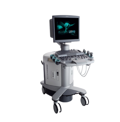
Ultrasonografi

General Imaging and Screening
Ultrasonography is a safe and non-invasive diagnostic method that uses high-frequency sound waves to visualize the internal structures of the body. Due to its lack of radiation, ease of application, and broad range of use, it is one of the most preferred imaging methods across nearly all medical specialties. It plays a key role in both diagnosis and follow-up, from pregnancy monitoring to the evaluation of internal organs.
Technological Infrastructure and Application
The ultrasound device transmits high-frequency sound waves through a handheld probe. These sound waves reflect back from body tissues, and the returning echoes are converted into real-time images by the device.
With Color Doppler ultrasonography, blood flow within vessels can be evaluated, and 3D/4D ultrasound technologies enable three-dimensional visualization of organs and structures. The procedure is completely painless, requires no special preparation, and provides immediate results — making ultrasonography one of the most widely used screening and diagnostic tools.
Advantages
- Safe imaging technique with no radiation exposure.
- Provides real-time visualization of internal organs.
- Fast, easy to perform, and delivers immediate results.
- Versatile method applicable to a wide range of medical conditions.
- Cost-effective and patient-friendly diagnostic option.
Clinical Applications
Ultrasonography is used across nearly all medical disciplines:
- Obstetrics and Gynecology: Pregnancy monitoring, uterus and ovary evaluations.
- Cardiology: Echocardiography for assessing cardiac structures and functions.
- General Surgery and Internal Medicine: Evaluation of organs such as the liver, kidneys, pancreas, gallbladder, and thyroid.
- Urology: Assessment of the prostate and bladder.
- Musculoskeletal System: Imaging of muscles, tendons, and joint pathologies.
- Emergency Medicine: Rapid detection of trauma, internal bleeding, or organ injuries.


